
Columbia University
Irving Medical Center
Neurological Institute
710 West 168th Street, 3rd floor
(212) 305-1818
TaubCONNECT Research Perspectives:
July 2021
1: Quantifying Age-Related Changes in Brain and Behavior: A Longitudinal Versus Cross-Sectional Approach
2: The Association Between Sex and Risk of Alzheimer's Disease in Adults with Down Syndrome
2: The Association Between Sex and Risk of Alzheimer's Disease in Adults with Down Syndrome
 |  | |
| Georgette Argiris, PhD | Christian Habeck, PhD |
Cognitive functions and their supporting neural mechanisms change across the lifespan. The knowledge about the nature and timing of this change is largely informed by cross-sectional studies, which have yielded variable results. For instance, some studies have observed lower brain activity in older compared to younger adults, suggesting a deficiency of processing, particularly when accompanied by lower behavioral performance, whereas others have observed higher activity, which has been linked to compensatory processing mechanisms. Since cross-sectional, age-related comparisons suffer from possible confounding variables, longitudinal studies to ascertain true aging-related effects are of paramount importance. Methodological challenges, however, such as demands of experimental design and participant attrition, have thwarted progress. Furthermore, of the longitudinal-aging studies to date, very few have comprehensively addressed a wide breadth of cognitive abilities.
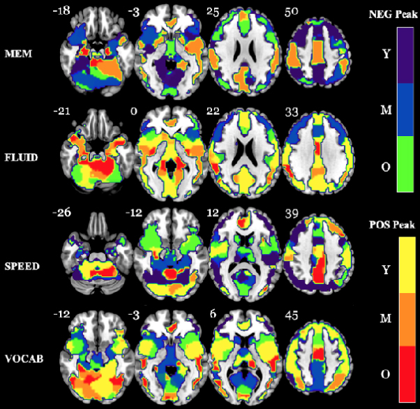
Figure. Longitudinal domain maps depicting the age bracket in which peak negative or positive changes occurred across the entire lifespan change curve. Regions-of-interest (ROIs), centered on each voxel in the brain, were created and the Gaussian age kernel was applied to each region. For each domain, we located the overall peak change value, irrespective of sign, across the entire change curve (essentially the maximum absolute value). The center voxel of each ROI was then color-coded depending on in which age bracket the peak was located and whether it was a positive or negative peak (see color bar to the right of the figure). The number next to each brain slice indicates the z-coordinate. NOTE: Slices are mirror-flopped where the right hemisphere is expressed on the left side. Y= Young; M= Middle Age; O= Ol
In the present study by Argiris et al., we employed in-scanner task-based functional magnetic resonance imaging to investigate both cross-sectional approximations of change versus actual longitudinal measurements of change in neural activation and behavioral performance across the lifespan (i.e., 20-80 years), in the Reference Ability Neural Network (RANN) cohort. Cognitively healthy participants were tested at two time points, a baseline assessment and again at 5-year follow-up, on a battery of tests relating to each of four cognitive reference abilities: Memory, Fluid Reasoning, Perceptual Speed, and Vocabulary. Using Gaussian Kernels at different ages, we generated “templates” of change, which is a concept borrowed from the MRI 4D atlas literature, where attention has been given to quantifying continuous lifespan changes. The merit of such an approach lies in refraining from overly restrictive statistical models, which often carry several assumptions imposing age-related linear effects.
We then compared (1) the change templates coming from cross-sectional approximations of age-related comparisons to (2) genuine longitudinal change templates in three discrete age brackets- young (20-40 years), middle (41-60 years), and old (61-80 years) age. We elucidated similarities and discrepancies of the two methods, with further probing of longitudinal change characteristics, such as peaks and fluctuations of change. Unsurprisingly, as recently reported in eNeuro, our results revealed several brain regions where cross-sectional and longitudinal measurements in each cognitive domain and age bracket differed markedly. Further, behavioral comparisons between cross-sectional and longitudinal measurements showed differences in change curves for all four domains, with processing speed displaying the steepest declines starting around 35 years of age, which was evident only in the longitudinal measurements. When focusing only on longitudinal measurements of neural change, we found more posterior brain regions that displayed peak activation increases, particularly for the old age bracket, with more frontal regions displaying peak activation decreases; this finding was generalizable across analysis types and cognitive domains. This is a notable and surprising result, given the prior support for age-related frontal increases, based on cross-sectional approximations. Our findings encourage greater focus on longitudinal measurements of aging-related changes, and show appreciable differences from possibly misleading cross-sectional approximations.
Georgette Argiris, PhD
Postdoctoral Research Scientist
gla2116@cumc.columbia.edu
Christian Habeck, PhD
Professor of Neurological Sciences (in Neurology and the Taub Institute) at CUMC
ch629@cumc.columbia.edu
The Association between Sex and Risk of Alzheimer's Disease in Adults with Down Syndrome
 |  | |
| Pooja Mhatre, MPH | Nicole Schupf, PhD |
Studies in the typically developing population have found that older age and the presence of an APOE-ε4 allele are the strongest factors increasing risk for Alzheimer’s disease (AD). There is some evidence that women appear to be at greater risk for developing AD compared with men, independent of the longer life expectancy for women. However, there is inconsistency in the literature, with most studies reporting higher risk in women, some reporting no difference in risk for AD by sex, and few studies reporting higher risk in men. Sex differences in the risk of AD in adults with Down syndrome (DS)—a population at high risk for AD related to triplication and overexpression of the gene for APP, located on chromosome 21—have not been extensively investigated. Similar to the typically developing population, existing studies have found conflicting results, with different studies showing onset for women being earlier, later, or no different than that for men.
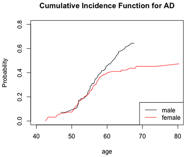
Figure. Cumulative incidence of AD by sex.
In the current study, together with first author and Mailman alumni '18 Pooja Mhatre, MPH, we examined the effect of sex on the risk of AD in adults with DS, adjusted for covariates. Adults with DS were assessed longitudinally for the development of AD. Competing risk survival analyses were used to determine the effect of sex alone and after adjustment for APOE-ε4 status, ethnicity, and level of intellectual disability (ID). As recently reported in the Journal of Clinical Medicine, we found that sex differences were significant only in adults over 60 years of age, where men with DS were 6.32 (95% CI: 2.11–18.96, p < 0.001) times more likely to develop AD compared with age-matched women with DS. Thus, as in the typically developing population, there is an age-associated effect of sex on the risk of AD, but there is higher age related risk in men than in women with DS. It will be important to examine if sex differences mediate the effects of other factors, such as comorbidities, metabolites, and genetic markers, on the risk of AD in this population.
Nicole Schupf, PhD
Professor of Epidemiology (in Neurology, Psychiatry, the Gertrude H. Sergievsky Center, and the Taub Institute for Research on Alzheimer's Disease and the Aging Brain)
ns24@cumc.columbia.edu
Contact Us
630 West 168th Street
P&S Box 16
New York, NY 10032
Phone: 212 305-1818
Fax: 212 342-2849
Email: taubinstitute@columbia.edu
Brain Donation
Our ability to understand Alzheimer's disease is dependent upon studying brain tissue.
For more information, please contact Donovan Laing at (212) 305-9086 dal2190@cumc.columbia.edu
This website uses cookies as well as similar tools and technologies to understand visitors' experiences. By continuing to use this website, you consent to Columbia University's usage of cookies and similar technologies, in accordance with the Columbia University Website Cookie Notice.