
Columbia University
Irving Medical Center
Neurological Institute
710 West 168th Street, 3rd floor
(212) 305-1818
TaubCONNECT Research Perspectives:
April 2020
1: » Cortical Thickness and its Associations with Age, Total Cognition and Education Across the Adult Lifespan
2: » Structural Brain Changes in Pre-Clinical FTD MAPT Mutation Carriers
3: » Phospholipase D1 Ablation Disrupts Mouse Longitudinal Hippocampal Axis Organization and Functioning
2: » Structural Brain Changes in Pre-Clinical FTD MAPT Mutation Carriers
3: » Phospholipase D1 Ablation Disrupts Mouse Longitudinal Hippocampal Axis Organization and Functioning
 |  | |
| Christian Habeck, PhD | Yunglin Gazes, PhD |
The importance of early-life education for cognition, health outcomes, and mortality has been well established. By its nature of being experienced in a sustained manner early in life, education (i.e., primary, secondary, and tertiary schooling) could be seen as a powerful intervention with likely beneficial effects for brain health even in mid- and late-life, regardless of overall cognitive status. In the current study, published recently in PLOS ONE, Drs. Christian Habeck, Yunglin Gazes, and colleagues from the Cognitive Neuroscience Division sought to identify univariate cortical-thickness patterns underlying education and general intelligence in 353 individuals aged 40 to 80.
As expected, people with longer education showed better global cognition and older people showed worse global cognition, despite a confounding positive relationship between age and education. However, the results of correlational analyses could not identify statistically robust associations between education and regional cortical thickness that went beyond associations with age, global cognition, and gender: even after statistically equating for these variables, people with longer education did NOT show correspondingly thicker cortices. Only when implementing more rigorous statistical controls that removed the associations of age, gender, and global cognition from both education and cortical thickness prior to performing correlations could a distributed education-related pattern of cortical thickness be obtained. This means concretely that the degree to which participants manifested the derived adjusted thickness pattern predicted their adjusted education duration (Figure 3, right panel). This pattern revealed that participants with higher education had relatively thicker cortices in right cingulate-cortex areas, while showing relatively thinner cortices in right-temporal areas.
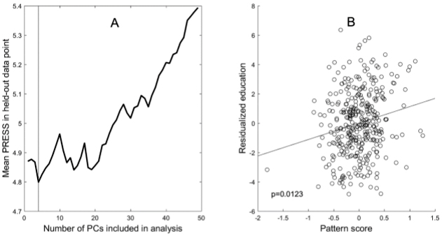 Figure 3: Results of the adjusted multivariate analysis to derive an education-related cortical-thickness pattern.
Panel A (left) shows the leave-one-out cross validation results which yield minimum mean PRESS for 4 Principal Components. Panel B (right) shows the relationship between subject scores of the derived pattern and the residualized education variable.
|
This study reaffirms the importance of early-life education for global cognition in mid- and late-life, but demonstrates the difficulty of finding corresponding associations with measures of brain structural health that are not already captured by global cognition.
Christian G. Habeck, PhD
Associate Professor of Neuroimaging (in Neurology and in the Taub Institute) at the CUMC
ch629@cumc.columbia.edu
Structural Brain Changes in Pre-Clinical FTD MAPT Mutation Carriers
 |  |  | ||
| Clara Domínguez-Vivero, MD | Edward D. Huey, MD | Stephanie Cosentino, PhD |
Frontotemporal dementia (FTD) is the second most common cause of early-onset neurodegenerative dementia. As with other neurodegenerative disorders, neuroimaging has been explored as a potential biomarker to identify patients in initial phases of FTD and to measure biological change over time. However, once subjects meet full criteria for the FTD diagnosis, structural changes are generally widespread.
Mutations in the microtubule-associated protein tau (MAPT) gene are among those identified in FTD. Studies specifically addressing preclinical neuroimaging features in MAPT mutation carriers may allow for characterization of the earliest neuroimaging changes in disease, but such studies are both scarce and inconsistent. Taub faculty members Dr. Stephanie Cosentino and Dr. Ted Huey established a longitudinal research protocol of a broadly phenotyped group of MAPT mutation carriers and familial matched controls. Thus, Drs. Huey and Cosentino, together with their research team including former postdoctoral fellow and first author Dr. Clara Dominguez-Vivero, recruited participants from this unique, well characterized population, in order to compare cortical and subcortical gray matter volumes, as well as white matter hyperintensities and tract-integrity, between MAPT mutation carriers and demographically-matched familial controls.
As recently reported in the Journal of Alzheimer’s Disease, they found that volumes of five cortical and subcortical areas were strongly correlated with mutation status: temporal lobe (left amygdala, left temporal pole), cingulate cortex (left rostral anterior cingulate gyrus, right posterior cingulate), and the lingual gyrus in the occipital lobe. They did not find significant differences in whole brain volume, white matter hyperintensities volume, and white matter integrity. These findings by Dominguez-Vivero et al. suggest the temporal lobe, cingulate cortex, and lingual gyrus to be early targets of the disease that may serve as neuroimaging biomarkers for FTD prior to overt symptom onset.
Stephanie Cosentino, PhD
Associate Professor of Neuropsychology (in Neurology, the Gertrude H. Sergievsky Center and the Taub Institute for Research on Alzheimer's Disease and the Aging Brain)
sc2460@cumc.columbia.edu
Edward D. Huey, MD
Associate Professor of Psychiatry and Neurology (in the Taub Institute for Research on Alzheimer's Disease and the Aging Brain)
edh2126@cumc.columbia.edu
Phospholipase D1 Ablation Disrupts Mouse Longitudinal Hippocampal Axis Organization and Functioning
 |  | |
| Tiago Gil Oliveira MD, PhD | Gilbert Di Paolo, PhD |
The hippocampus is a brain structure fundamental for learning and memory, and despite maintaining its cross-sectional architecture along its longitudinal axis, anatomical, gene expression and functional studies suggest an organized gradient that segregates in rodents dorsal and ventral poles.
In this study, an international team of researchers, led by Dr. Tiago Gil Oliveira, a former PhD student at Taub and now Assistant Professor at University of Minho in Portugal, in collaboration with Dr. Gilbert Di Paolo while he was a Taub faculty, showed that the lipid modulating enzyme, phospholipase D1 (PLD1), is involved in the organization and functioning of the hippocampus along its longitudinal axis. They followed up their previous observations from an unbiased lipidomic analysis that showed a dorsal-ventral hippocampal gradient of the signaling lipid, phosphatidic acid (PA), the product of PLD.
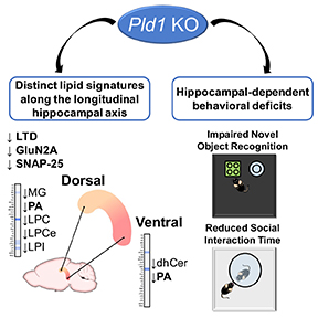
As published in Cell Reports by Luísa Santa-Marinha et al, the authors used genetic models of PLD1 and PLD2 ablation, the two canonical mammalian PLD enzymes, and observed that, comparatively to PLD2, PLD1 contributed the most to total hippocampal levels of PA. Further analysis showed that PLD1 knock-out mice present deficits in object recognition and social exploration, reduction in long-term depression induction, and decreased levels of the synaptic proteins GluN2A and SNAP-25 in the dorsal hippocampus.
Tiago Gil Oliveira, MD, PhD
Assistant Professor, School of Medicine, University of Minho
tiago@med.uminho.pt
Contact Us
630 West 168th Street
P&S Box 16
New York, NY 10032
Phone: 212 305-1818
Fax: 212 342-2849
Email: taubinstitute@columbia.edu
Brain Donation
Our ability to understand Alzheimer's disease is dependent upon studying brain tissue.
For more information, please contact Donovan Laing at (212) 305-9086 dal2190@cumc.columbia.edu
This website uses cookies as well as similar tools and technologies to understand visitors' experiences. By continuing to use this website, you consent to Columbia University's usage of cookies and similar technologies, in accordance with the Columbia University Website Cookie Notice.