
Columbia University
Irving Medical Center
Neurological Institute
710 West 168th Street, 3rd floor
(212) 305-1818
Featured Research
In the Lab:
Wassim Elyaman, PhD
Wassim Elyaman, PhD
Although historically considered an immune-privileged site, the central nervous system (CNS) is now known to be involved in lymphatic drainage and actively communicates with the peripheral immune system to regulate immune responses, both centrally and peripherally. My laboratory is interested in understanding the influence of aging on the immune system, also known as “immunosenescence,” and its consequence on the development of neurodegenerative diseases such as Alzheimer’s disease (AD) and Parkinson’s disease (PD). My interest in studying peripheral immune cells in these debilitating diseases stems from my previous training, first as a research fellow and later as a faculty in the Department of Neurology at Brigham and Women’s Hospital, where I studied molecular pathways leading to adaptive immune responses in autoimmune diseases, particularly multiple sclerosis (MS).
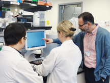
From left: Research Fellow Chunsheng (Kevin) Ruan, PhD; Research Assistant Alex Kroshilina, BS; and Wassim Elyaman, PhD.
As co-director of basic research in the Center for Translational and Computational Neuroimmunology, (CTCN; directed by Taub faculty member Dr. Philip L. De Jager), I work closely with clinical immunologists, geneticists, computational biologists, and neurologists to leverage human transcriptomics and epigenomics data, generated using patient brain autopsies and biopsies, to identify novel immune molecules involved in neurodegeneration. In an ongoing collaboration with Taub faculty member Dr. Elizabeth (Betsy) Bradshaw, we are generating a biobank of T cell clones from the brain of AD and PD autopsies. Additionally, we are sequencing microglia MHC-bound peptides to identify self and non-self peptides that are being presented to T cells in AD and PD patients. This exciting research avenue will lead to the identification of intrinsic (auto-antigens) and extrinsic (environmental) factors that may have therapeutic potential.
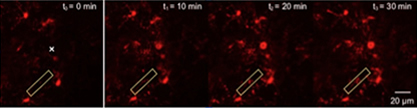
Figure 1. Time-lapse two-photon imaging of microglia during injury using the TMEM119-tdTomato mice.
In order to further characterize the microglia-T cell crosstalk in vivo and test lead immunomodulators, my laboratory has developed novel in vitro and in vivo reporter mouse models on the AD and the PD backgrounds. Indeed, we have engineered a dual reporter mouse model expressing microglia-specific marker TMEM119, tagged with a red fluorescent protein (tdTomato), and Foxp3, a marker of regulatory T cells conjugated with a green fluorescent protein (GFP). In collaboration with Dr. Guang Yang in the Department of Anesthesiology, we are analyzing the interaction between microglia and regulatory T cells in AD and PD mice using two-photon microscope (Figure 1). Moreover, we have generated another dual reporter mouse model to distinguish between CNS-resident innate immune cells (microglia expressing TMEM119-tdTomato) and infiltrating innate immune cells (macrophages expressing CX3CR1-GFP) in genetic mouse models of AD and PD.
Finally, my laboratory is interested in identifying the molecular pathways that connect genetic risk variants associated with MS with immune targets leading to increased susceptibility to develop an autoimmune response. In collaboration with Dr. Philip De Jager, we are utilizing omics data sets to translate genetic association into functional outcome using human and murine systems that will potentially lead to druggable targets.
Wassim Elyaman, PhD
Assistant Professor of Neurological Sciences (in Neurology, the Taub Institute and the Institute for Genomic Medicine)
we2152@cumc.columbia.edu
https://www.columbiactcn.org
Contact Us
630 West 168th Street
P&S Box 16
New York, NY 10032
Phone: 212 305-1818
Fax: 212 342-2849
Email: taubinstitute@columbia.edu
Brain Donation
Our ability to understand Alzheimer's disease is dependent upon studying brain tissue.
For more information, please contact Donovan Laing at (212) 305-9086 dal2190@cumc.columbia.edu
This website uses cookies as well as similar tools and technologies to understand visitors' experiences. By continuing to use this website, you consent to Columbia University's usage of cookies and similar technologies, in accordance with the Columbia University Website Cookie Notice.